
Introduction
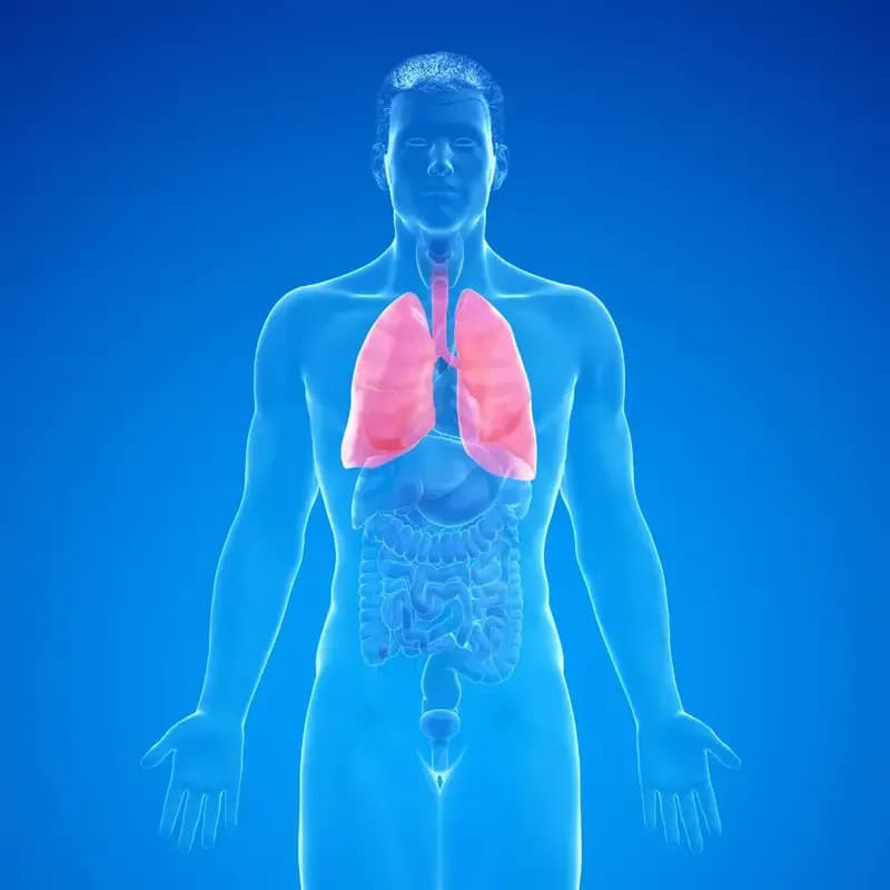
Cystic lung disease refers to a group of conditions characterized by the presence of multiple cysts or air-filled sacs in the lungs. These cysts can vary in size and number and may develop due to various underlying causes, including genetic mutations, infections, inflammatory conditions, and environmental factors. Cystic lung disease can affect lung function and respiratory health, leading to symptoms such as shortness of breath, coughing, and recurrent respiratory infections.
Types of Cystic Lung Disease
There are several types of cystic lung disease, each with its own distinct characteristics and underlying causes. Some common types include lymphangioleiomyomatosis (LAM), pulmonary Langerhans cell histiocytosis (PLCH), bronchiectasis, and congenital cystic lung diseases such as congenital pulmonary airway malformation (CPAM) and bronchogenic cysts. Understanding the specific type of cystic lung disease is essential for determining the appropriate treatment approach.
Causes and Risk Factors
The exact cause of cystic lung disease may vary depending on the specific type of condition. In some cases, cystic lung disease may result from genetic mutations or inherited disorders that affect lung development and function. Other risk factors for cystic lung disease may include exposure to environmental toxins, smoking, respiratory infections, and certain medical conditions such as rheumatoid arthritis and sarcoidosis.
Symptoms and Clinical Manifestations
The symptoms of cystic lung disease can vary widely depending on the size, location, and number of cysts present in the lungs. Common symptoms may include progressive shortness of breath, chronic cough, wheezing, chest pain, fatigue, and recurrent respiratory infections. In severe cases, cystic lung disease can lead to respiratory failure and other complications that may require medical intervention.
Diagnosis and Evaluation
Diagnosing cystic lung disease typically involves a combination of medical history review, physical examination, imaging studies (such as chest X-rays, CT scans, or MRI), pulmonary function tests, and sometimes biopsy or genetic testing. These diagnostic tests help healthcare providers assess the extent of lung involvement, determine the underlying cause of the cystic lung disease, and develop an appropriate treatment plan.
Treatment Options
The treatment approach for cystic lung disease depends on several factors, including the type and severity of the condition, the presence of symptoms, and the individual's overall health status. Treatment options may include medications to manage symptoms and slow disease progression, pulmonary rehabilitation to improve lung function and quality of life, oxygen therapy, and in some cases, surgical intervention to remove cysts or repair damaged lung tissue.
Lifestyle Management and Supportive Care
In addition to medical treatment, lifestyle modifications and supportive care measures can help individuals with cystic lung disease manage their symptoms and improve respiratory health. These may include smoking cessation, avoiding exposure to environmental pollutants and respiratory irritants, maintaining a healthy weight, staying physically active, and following a nutritious diet rich in fruits, vegetables, and whole grains.
Prognosis and Outlook
The prognosis for individuals with cystic lung disease can vary depending on the specific type and severity of the condition, as well as the effectiveness of treatment interventions. While some forms of cystic lung disease may have a relatively benign course, others may progress over time and lead to significant respiratory impairment and complications. Early diagnosis, comprehensive medical care, and lifestyle modifications can help improve outcomes and enhance quality of life for individuals living with cystic lung disease.
Conclusion
Cystic lung disease encompasses a diverse group of conditions characterized by the presence of cysts or air-filled sacs in the lungs. While the exact cause and prognosis may vary depending on the specific type of cystic lung disease, early recognition and appropriate management are essential for optimizing outcomes and improving respiratory health. With advancements in diagnostic techniques and treatment modalities, individuals with cystic lung disease can benefit from tailored interventions aimed at controlling symptoms, slowing disease progression, and enhancing overall quality of life. By working closely with healthcare providers and adopting healthy lifestyle habits, individuals can effectively manage cystic lung disease and minimize its impact on daily functioning and long-term health. Continued research and innovation in the field of respiratory medicine hold promise for further improving the diagnosis, treatment, and management of cystic lung disease in the future.









Comments (0)