

Introduction
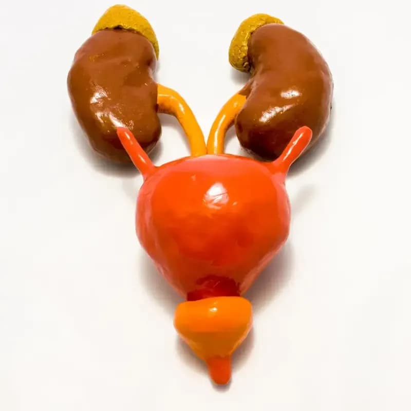
A trabeculated bladder is a condition characterized by thickening and irregularity of the bladder wall. This condition often results from prolonged obstruction of urinary flow, leading to changes in the bladder's structure and function. In this article, we will explore the causes, symptoms, and treatment options for a trabeculated bladder, and provide tips for managing this condition effectively.
Causes of Trabeculated Bladder
Trabeculated bladder can be caused by several factors, most commonly due to chronic bladder outlet obstruction. This obstruction can be a result of conditions such as benign prostatic hyperplasia (BPH) in men, urethral stricture, or bladder neck contracture. Prolonged urinary retention forces the bladder muscles to work harder to expel urine, leading to hypertrophy and trabeculation. Other causes include neurogenic bladder, where nerve damage affects bladder function, and chronic inflammation or infection of the bladder.
Symptoms of Trabeculated Bladder
The symptoms of a trabeculated bladder can vary depending on the severity of the condition and the underlying cause. Common symptoms include:
Increased frequency of urination, often with a sense of urgency, is a common symptom. Patients may feel the need to urinate frequently throughout the day and night.
Frequent Urination
A sensation of incomplete bladder emptying after urination can occur. This can lead to repeated trips to the bathroom.
Incomplete Emptying
A weak or intermittent urine stream is another common symptom. Patients may find it difficult to start urinating or maintain a steady flow.
Weak Urine Stream
Straining or pushing to initiate urination can be a symptom of trabeculated bladder. This is often due to the increased effort required by the bladder muscles.
Straining to Urinate
In severe cases, patients may experience urinary retention, where the bladder is unable to empty completely, leading to discomfort and potential complications.
Urinary Retention
Diagnosing Trabeculated Bladder
Diagnosis of a trabeculated bladder typically involves a combination of patient history, physical examination, and diagnostic tests. These may include:
Ultrasound imaging can help visualize the bladder wall and detect signs of trabeculation. It is a non-invasive and commonly used diagnostic tool.
Ultrasound
A cystoscopy involves inserting a thin, flexible tube with a camera into the bladder through the urethra. This allows direct visualization of the bladder wall and any structural abnormalities.
Cystoscopy
Urodynamic studies measure the pressure and flow of urine in the bladder and urethra. These tests can assess how well the bladder and urethra are storing and releasing urine.
Urodynamic Tests
In some cases, advanced imaging techniques like MRI or CT scans may be used to provide detailed images of the bladder and surrounding structures.
MRI or CT Scan
Treatment Options for Trabeculated Bladder
Treatment for a trabeculated bladder depends on the underlying cause and severity of the condition. Options may include:
Medications such as alpha-blockers or 5-alpha reductase inhibitors can help relieve symptoms by reducing bladder outlet obstruction and improving urine flow.
Medications
Intermittent or indwelling catheterization may be necessary to ensure complete bladder emptying in cases of severe urinary retention.
Catheterization
Surgery may be required to correct underlying conditions such as BPH, urethral stricture, or bladder neck contracture. Procedures like transurethral resection of the prostate (TURP) or urethroplasty can help alleviate obstruction.
Surgical Intervention
Lifestyle changes, including fluid management, bladder training, and pelvic floor exercises, can help manage symptoms and improve bladder function.
Lifestyle Modifications
Preventing Trabeculated Bladder
While it may not always be possible to prevent a trabeculated bladder, certain measures can help reduce the risk:
Regular medical check-ups can help detect and manage conditions like BPH or urinary tract infections early, preventing complications that can lead to bladder trabeculation.
Regular Check-Ups
Maintaining a healthy lifestyle with a balanced diet, regular exercise, and adequate hydration can support overall urinary health.
Healthy Lifestyle
Practicing good bladder habits, such as avoiding holding urine for prolonged periods and ensuring complete bladder emptying, can help maintain bladder health.
Bladder Health
Conclusion
A trabeculated bladder is a condition that requires proper diagnosis and management to prevent complications and improve quality of life. By understanding the causes, symptoms, and treatment options, patients and healthcare providers can work together to develop an effective management plan. Regular monitoring and lifestyle modifications can also play a significant role in maintaining bladder health and preventing further complications. If you experience symptoms of a trabeculated bladder, consult with a healthcare professional for appropriate evaluation and treatment.







Comments (0)