Conjunctivitis (Eye Flu): A Detailed Guide
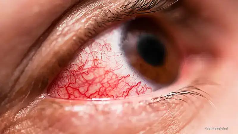
Conjunctivitis, commonly known as eye flu, is an inflammation of the conjunctiva that causes redness, irritation, and discomfort.

Eye emergencies require a different response from most medical problems — some need action within minutes (chemical splash, retinal artery occlusion), others within hours (retinal detachment, acute glaucoma), and still others within days. The common thread is that delays cause irreversible harm: vision lost to a retinal detachment not repaired within hours, vision destroyed by a chemical burn not irrigated immediately, optic nerve permanently damaged in an acute glaucoma attack not treated the same day. In India, awareness of eye emergencies is low — patients with sudden vision loss often try home remedies for days before attending a hospital. This hub focuses on recognising the genuine eye emergency, knowing the first-aid response (especially for chemical injuries), and getting to the right place fast.
Last reviewed: 11 August 2026
Conjunctivitis, commonly known as eye flu, is an inflammation of the conjunctiva that causes redness, irritation, and discomfort.

Eye flu, commonly known as viral conjunctivitis, is an inflammation of the conjunctiva, the thin, clear tissue covering the white part of the eye and the inside of the eyelids.
Expert answers from our medical team
Retinal detachment occurs when the retina separates from the back of the eye — it is painless but causes characteristic symptoms: a sudden shower of new floaters (dark spots or cobweb shapes that appear suddenly in large numbers), flashes of light (photopsia) especially in the peripheral vision, and — if the detachment has reached the macula — a dark curtain or shadow encroaching from one side of vision. Any new sudden floaters with flashing lights needs urgent ophthalmology assessment — same day if possible. A peripheral retinal tear without full detachment can be treated with laser (photocoagulation) or cryotherapy to seal it before it progresses. Once the retina detaches completely, surgery (vitrectomy or scleral buckling) is needed and visual outcome depends on how quickly it is repaired — macula-on detachments repaired within 24 hours have significantly better outcomes.
Acute angle-closure glaucoma is an ocular emergency — nothing like the silent, gradual open-angle glaucoma. It occurs when the drainage angle of the eye suddenly closes, causing intraocular pressure to spike dramatically (sometimes above 60-70 mmHg, versus normal of 10-21). Symptoms: severe eye pain (often described as the worst pain the person has felt), headache, nausea and vomiting, blurred vision, and coloured halos around lights. The eye is red and the cornea may appear steamy or hazy. Untreated, it causes permanent severe vision loss within hours. Treatment: emergency pressure-lowering eye drops, intravenous acetazolamide, and urgent laser iridotomy to re-open the drainage angle. It is most common in hyperopic (long-sighted) people, women, those with shallow anterior chambers — and can be precipitated by dim light, certain medications, or stress.
Immediate, copious irrigation is the single most important first aid measure — every minute of delay allows more chemical penetration. Flush the eye with the nearest available water (tap water, bottled water, saline) continuously for at least 20 minutes. Hold the eye open during irrigation — use your fingers if needed. Remove contact lenses if present before or during irrigation. Don't waste time finding a special solution, calling a doctor, or putting on protective gear before starting — start flushing immediately. Alkalis (cement, lime, bleach, ammonia) penetrate more deeply and cause more severe damage than acids — but all chemical injuries need the same immediate response. After 20 minutes of irrigation, go directly to an emergency eye unit. Do not patch the eye. Bring the chemical bottle or label if possible — the pH guides treatment. Time from injury to irrigation is the primary determinant of outcome.
Sudden vision loss is always an emergency. Possible causes include: retinal artery occlusion (a 'stroke' of the retinal artery — treatable with interventions if presented within hours), retinal detachment, acute glaucoma attack, vitreous haemorrhage (bleeding inside the eye, often from diabetic retinopathy), optic neuritis (inflammation of the optic nerve, associated with multiple sclerosis), and stroke affecting the visual cortex. Some causes (retinal artery occlusion) have treatment windows measured in hours. Others (retinal detachment) in hours to days. Do not try home remedies, do not 'wait and see if it gets better,' and do not accept 'come next week' from any clinic. Go to a hospital with an ophthalmologist on call immediately — call ahead to confirm eye emergency cover. Describe the onset (sudden or gradual), whether one or both eyes, and any associated symptoms (pain, headache, neurological signs).
Superficial foreign bodies (dust, grit, eyelash): blink repeatedly, pull the upper eyelid over the lower lash and hold briefly (lower lashes may dislodge the particle), or irrigate with clean water or saline. If the object is visible under the eyelid, a clean damp cotton bud tip can gently lift it. Never rub the eye — this embeds the particle and scratches the cornea. If the particle doesn't come out easily, cover the eye and seek clinic attention. Don't attempt removal with sharp objects. Penetrating injury (metal fragment, nail, wire, wood splinter that may have entered the eye): this is a true emergency. Do not rub, do not press on the eye, do not try to remove the object. Cover with a rigid shield (a cup held over the eye without touching it), keep the person still and upright, and go directly to an emergency eye unit. Pressure on a penetrating injury can expel ocular contents.
Questions from our community members
Ask our medical community. Your question may help others with similar concerns.
Verified specialists across India