

Overview
Understanding Follicle Studies for Fertility
A follicle study, also known as follicular monitoring or follicle tracking, involves a series of ultrasound scans to observe the development of ovarian follicles throughout the menstrual cycle. This procedure is commonly used by doctors to assess ovulation, monitor treatments aimed at inducing ovulation, or to plan the timing of fertility treatments.
Many patients begin this process with a basic understanding that their doctor wants to “check the egg.” While this is a simplified explanation, the ultrasound actually focuses on the follicle, which is the fluid-filled sac within the ovary where an egg matures. The key information gathered comes from observing the follicle's growth, the condition of the uterine lining, and signs that ovulation may be occurring.
Preparing for Your Follicle Study
Know Your Cycle Day
The first day of your menstrual period is typically considered Day 1 of your cycle. Follicular monitoring is scheduled based on the expected development of the follicle, not just on appointment availability. The exact schedule can vary; some fertility clinics start with a baseline scan early in the cycle, followed by repeat scans around the anticipated fertile period. Others may begin monitoring around Day 10 to 12, especially during ovulation-induction treatments. Additional scans might be necessary depending on the findings.
If you have irregular cycles, your scan schedule may differ from someone with a standard 28-day cycle. Your doctor may adjust the timing based on your specific cycle and the treatment you are receiving. It's helpful to keep a record of your period's start date and bring details of any current medications, particularly fertility drugs like letrozole, clomiphene, or gonadotrophins, as these can influence the scan schedule.
Bladder Preparation
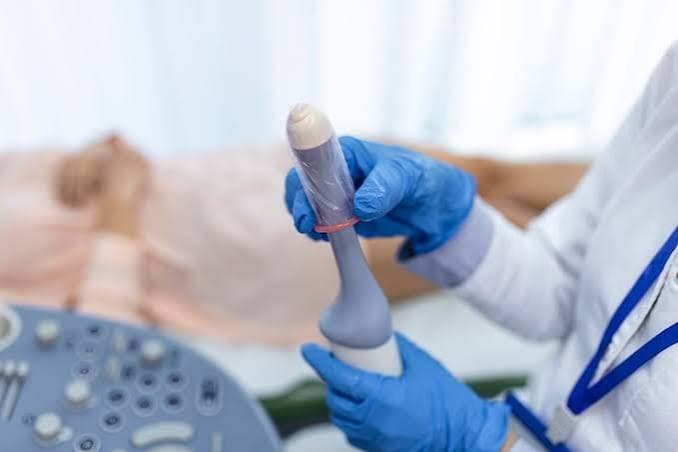
For follicular monitoring, a transvaginal ultrasound is usually performed, offering a clearer view of the ovaries and uterus compared to an abdominal scan. The ultrasound probe is gently inserted into the vagina, and the procedure is typically brief. Many patients are unaware that a full bladder is generally not required for this type of scan; in fact, fertility units often instruct patients to empty their bladder beforehand. Always follow the specific preparation instructions provided by your clinic, as practices can vary.
This ultrasound examination does not involve radiation and is considered a routine and generally well-tolerated procedure.
What Doctors Look for During the Scan
The scan aims to gather more information than just the presence of an egg.
- Follicle Development: Clinicians assess the number and size of developing follicles.
- Ovarian Appearance: The overall condition of the ovaries is examined.
- Uterine Lining: The thickness and appearance of the endometrium (the lining of the uterus) are evaluated.
As the cycle progresses, a dominant follicle may enlarge and move towards ovulation. It's a common misconception that an 18 mm measurement refers to the egg's size; this measurement actually indicates the size of the follicle itself. The egg, being microscopic, cannot be directly measured by routine ultrasound. While follicles often reach maturity around the middle of the cycle, the interpretation depends on the growth pattern, treatment being used, hormone levels, and signs of ovulation.
The Importance of Serial Scans
If your doctor requests a follow-up scan in a few days, it's usually to observe changes over time. A single ultrasound provides a snapshot, while a series of scans reveals growth patterns, the development of multiple follicles, and whether ovulation has occurred. This serial monitoring is especially crucial during ovulation-induction treatment, as responses to fertility medications can vary. It allows clinicians to adjust treatment and identify potential overstimulation.
Follicle Growth vs. Pregnancy
A well-developed follicle is a positive sign of ovarian response and approaching ovulation, but it does not guarantee pregnancy. Conception requires a complex sequence of events, including ovulation, fertilization in the fallopian tube, embryo development, and implantation in the uterus. A follicle study primarily provides information about follicular development and ovulation; other fertility factors may need separate assessment.
Interpreting Scan Results and Timing
Avoid making decisions about intercourse or treatment timing based solely on a single scan number. The appropriate timing for intercourse, ovulation-trigger injections, IUI, or other fertility procedures is determined by your individual treatment plan and the overall monitoring results. In stimulated cycles, ultrasound findings and sometimes hormone levels guide treatment timing. Always follow the specific instructions from your fertility team rather than comparing your measurements with online information.
Questions to Ask Your Clinician
To better understand your follicle study report, consider asking your clinician the following:
- How many developing follicles are present?
- Which follicle is the leading one?
- Is the follicle growing as expected for this stage of my cycle?
- What is the appearance of my uterine lining?
- Do I need another scan or a blood test?
- Does my response to fertility medication appear appropriate?
- When should we plan for intercourse or return for treatment?
Focusing on these questions can provide a clearer understanding than concentrating on a single number.
Context Matters: Treatment Goals
The significance of a follicle study is tied to its purpose. For those confirming natural ovulation, it verifies follicle development and egg release. In ovulation induction, it assesses ovarian response and checks for overstimulation. For assisted fertility treatments, it helps determine the optimal timing for procedures. Therefore, similar ultrasound measurements can lead to different advice based on individual treatment plans.
Key Takeaway
A follicle study is best viewed as a narrative unfolding over several scans, rather than a single data point. Before your appointment, be aware of your cycle day, bring your medication details, follow clinic instructions for bladder preparation, and understand that a transvaginal scan is common. During monitoring, inquire about follicle growth, the uterine lining, signs of ovulation, and the next steps. Most importantly, do not solely rely on follicle measurements to gauge the success of a fertility cycle; the scan provides one crucial piece of the fertility puzzle.



Comments (0)